


pCD2-hTL1A-Tg
Nomenclature
C57BL/6JSmo-Tgtn(pCD2-hTL1A)1Smoc
Cat. NO.
NM-TG-250279
Strain State
Sperm cryopreservation
Model Description
Validation Data

Fig.1 Detection of human TL1A expression in serum by ELISA (n=3, 6 weeks old).
Abbr. WT, wild type; N.D. not detected.
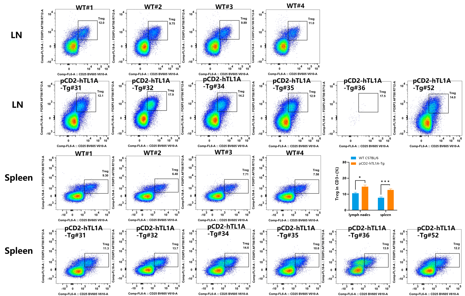
Fig.2 Detection of Treg expression on CD3+T cells in MLN and spleen of WT (C57BL/6) mice and pCD2-hTL1A-Tg mice by FACS.
In CD3+ T cells, compared with wild-type mice, the number of Treg cells in the spleen and MLN of pCD2-hTL1A-Tg mice was increased (n=4-6, 17 weeks old).
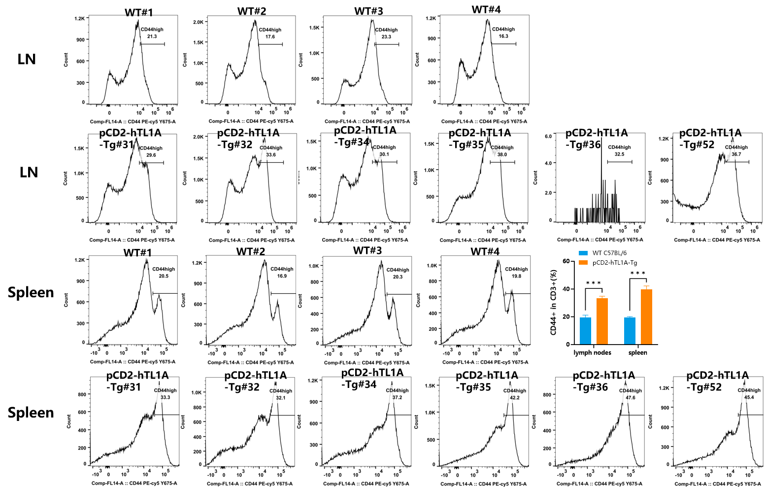
Fig.3 Detection of CD44 expression on CD3+T cells in MLN and spleen of WT (C57BL/6) mice and pCD2-hTL1A-Tg mice by FACS.
In CD3+ T cells, compared with wild-type mice, the number of CD44+ cells in the spleen and MLN of pCD2-hTL1A-Tg mice was increased (n=4-6, 17 weeks old).
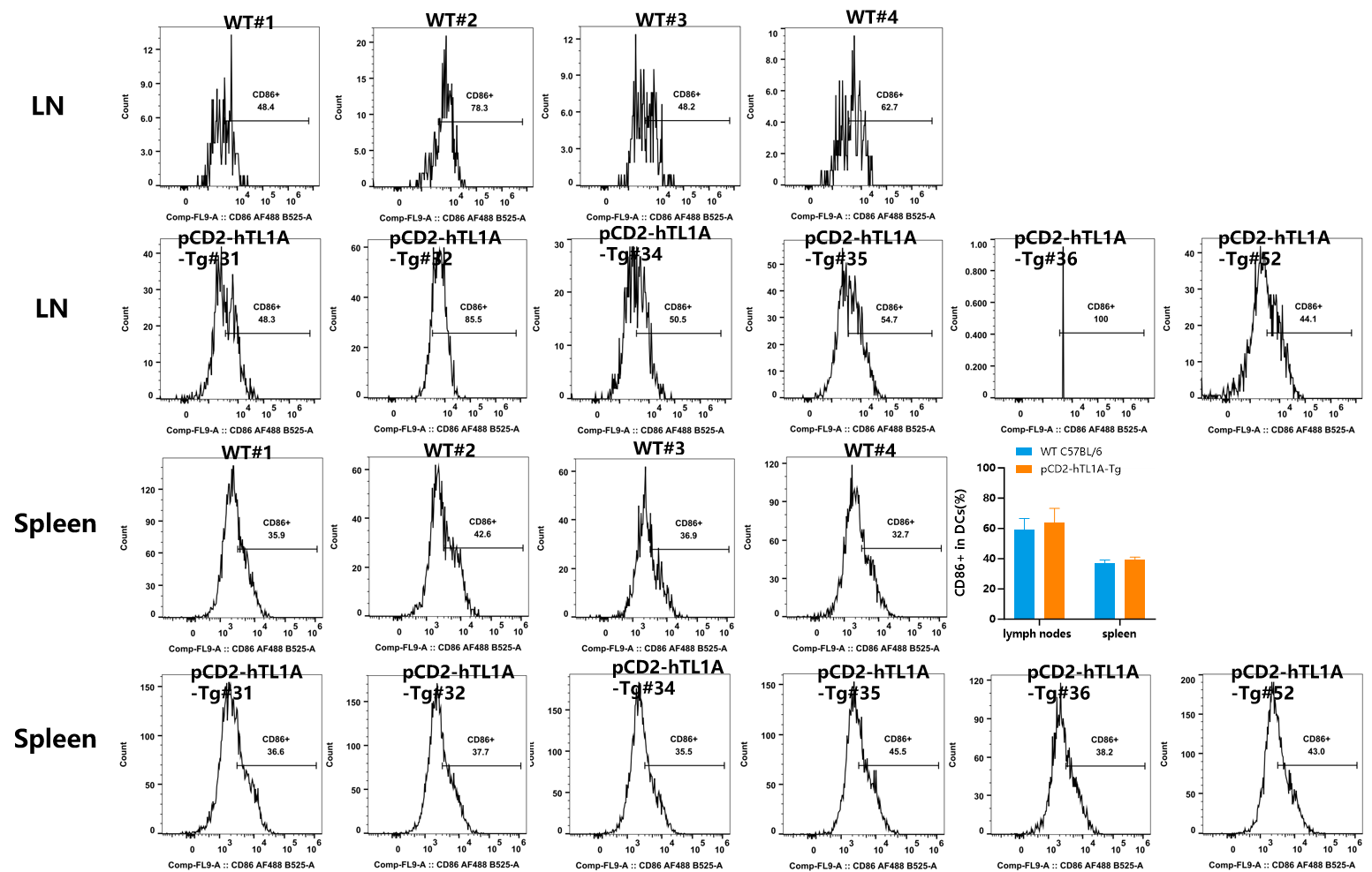
Fig.4 Detection of CD86 expression on CD11c+DCs in MLN and spleen of WT (C57BL/6) mice and pCD2-hTL1A-Tg mice by FACS.
In DCs cells, the number of CD86+ cells in the spleen and MLN of pCD2-hTL1A-Tg mice was similar to that of wild-type mice (n=4-6, 17 weeks old).
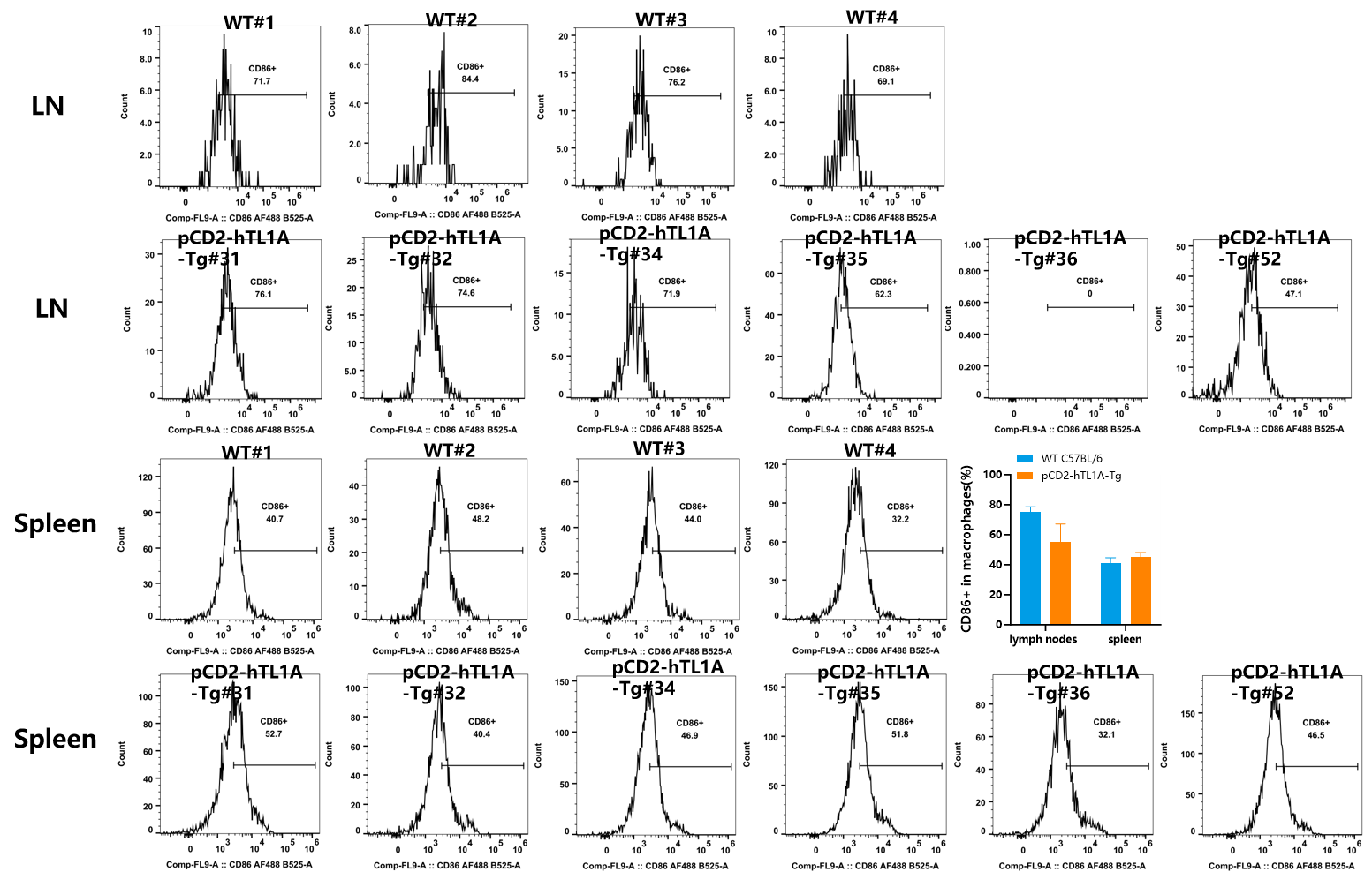
Fig.5 Detection of CD44 expression on macrophages in MLN and spleen of WT (C57BL/6) mice and pCD2-hTL1A-Tg mice by FACS(n=4-6, 17 weeks old).
In macrophages, the number of CD86+ cells in the spleen and MLN of pCD2-hTL1A-Tg mice was similar to that of wild-type mice.


Fig.6 Detection of IFN-γ expression on CD4+T cells in MLN and spleen of WT (C57BL/6) mice and pCD2-hTL1A-Tg mice by FACS.
In CD4+ T cells, compared with wild-type mice, the number of IFNγ+ cells in the spleen and MLN of pCD2-hTL1A-Tg mice was significantly increased (n=4-6, 17 weeks old).

Fig.7 Detection of IL-17A+ expression on CD4+T cells in MLN and spleen of WT (C57BL/6) mice and pCD2-hTL1A-Tg mice by FACS.
In CD4+ T cells, compared with wild-type mice, the number of IL-17A+ cells in the spleen and MLN of pCD2-hTL1A-Tg mice was significantly increased (n=4-6, 17 weeks old).

Fig.8 Detection of IL-4+ expression on CD4+T cells in MLN and spleen of WT (C57BL/6) mice and pCD2-hTL1A-Tg mice by FACS.
No IL-4+ cells were observed in the spleen and MLN of pCD2-hTL1A-Tg mice and wild-type mice (n=4-6, 17 weeks old).

Fig. 8 Phenotype of WT (C57BL/6) mice and pCD2-hTL1A-Tg mice (n=2-3, 17 weeks old). (A) Gross photograph; (B) Colon photo; (C) Body weight and intestine index.

Fig. 9 H&E staining of the small intestine and colon in pCD2-hTL1A-Tg mice (n=2-3, 17 weeks old).

Fig. 10 Representative masson's trichrome staining images of the small intestine and colon in pCD2-hTL1A-Tg mice. (n=2-3, 17 weeks old)
You may also like
Workshop:Progress and Advances in Preclinical immuno-Oncology Research
SMOC’s Annual Progress and Advances in Preclinical immuno-Oncology Research: The workshop is designed as a forum for ideas and opinions exchange on how to decrease the rate of clinical failures in oncology and immuno-oncology.
Learn moreShanghai Model Organisms Center Inc has licensed CRISPR-Cas9 technology from Broad Institute
On Dec 16, 2018, Broad Institute and Shanghai Model Organisms Center Inc (SMOC) has entered into a non-exclusive license agreement under which Broad has granted SMOC worldwide rights to commercialize a service platform for genetically modified mouse models under Broad's intellectual property.
Learn moreCustomizing Mouse Models: Delivering in 100 Days to Speed Up Your Research!
At GenoBioTX, we understand that the lengthy wait times for gene-modified mouse models can hinder your research progress. Traditional methods often require 6-9 months, leading to delays and increased costs. That’s why we’re thrilled to introduce our innovative service designed to streamline this process and deliver results faster.
Learn more
